| Phase Boundary Migration in DCB. |
Click image to enlarge (~1MB) |
|
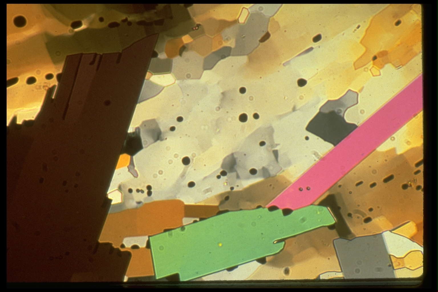
DBC has a monoclinic (alpha) structure stable at room temperature and a triclinic (beta) structure stable above 31°C. The two polymorphs have very similar densities (within 2%). This series of pictures was made with the motor on, but little or no deformation occurred in the field of view. The microstructural changes recorded were mainly induced by changing the temperature. The boundaries between alpha and beta grains are called PHASE BOUNDARIES, to distinguish them from GRAIN BOUNDARIES separating grains of the same phase. |
|
| 48. A
field of alpha grains and subgrains (white, grey, light brown, and
orange), in which three idioblastic porphyroblasts of the beta phase
have grown; the large dark brown grain at the left, a green grain, and
a pink one. The temperature of the sample is about 33°C. The field
of view is only about 400µm wide. The black blobs are vapor-filled holes that extend right through the sample. They represent vapor bubbles in the DCB melt from which the sample was initially grown. The holes serve as imperfect material markers. (They are capable of moving through the material to some extent. They can also grow, or shrink and vanish.) |
|
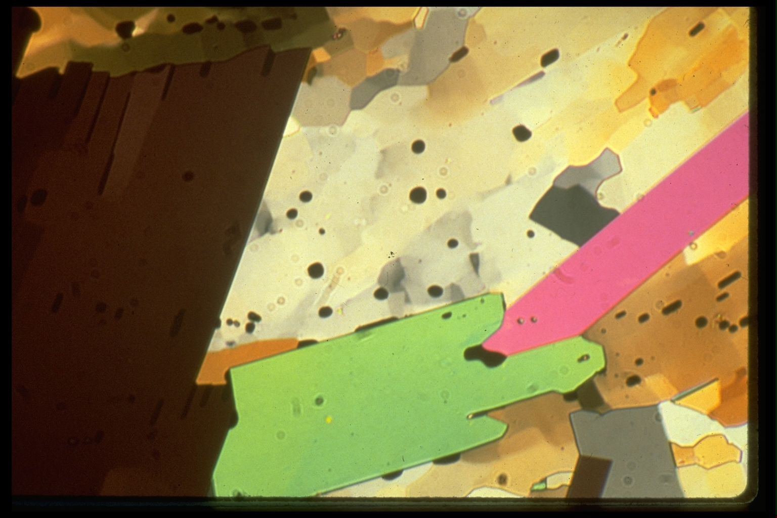
| 49. Ten minutes later. The temperature has been
increased and is now 42°C. All three beta grains have grown. See
how the boundary of the brown grain at (35,37) has overgrown grains and
subgrains in the alpha material without losing its straightness. It has
migrated at a uniform velocity to the right, regardless of the lattice
orientation of the unstable alpha material being replaced. Use the holes as markers in this photo and the last one, to work out whether both boundaries of the pink crystal are migrating at the same rate. How does the asymmetric growth of the pink grain square with behavior of the brown grain's boundary just observed? Is there a problem here? (ans) Which boundary of the green grain has migrated faster, the top or the bottom boundary? Notice the interaction of the holes and the top boundary of the green grain. The holes, from (40,55) to (55,50) in the previous image, appear to have been swept along with the boundary and partly amalgamated into the elongate hole now at (52,45). The hole originally at (59,50) on the other hand, appears to have been swept for a while, and then poikoblastically incorporated in the green grain, at its present location (62,43). What determines whether a hole is pushed ahead of the phase boundary or overgrown by it? The fact that the green grain seems to have grown as fast where it is against a hole (DCB vapor) as where it is against a crystal, can be interpreted to indicate that there is an invisibly thin vapor layer between alpha DCB and these beta porphyroblasts everywhere. Do you see why? See the brown beta porphyroblast replacing the grey alpha grain at (80,65). This is actually another arm of the large brown beta crystal at (20,40). |
|
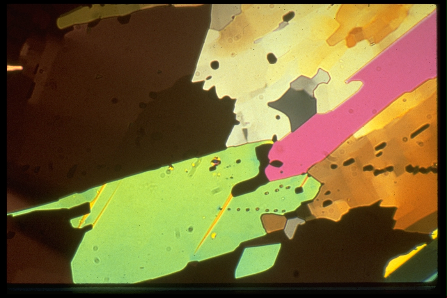
| 50. Nine minutes after the last slide. The heater
is off and the temperature is going down, but it is still above the
beta/alpha transition. More growth of the brown grain is seen, but
locally it is being replaced by the green one. Why should one
porphyroblast replace another when both are nominally stable? (ans) More growth of the pink grain, this time on
its other side. The next photo was taken three quarters of an hour after this one, by which time the temperature of the sample had fallen to about 26°C (5° below the nominal beta/alpha transition temperature). See if you can predict the kinds of microstructural changes to be expected. |
|
| 51. The unstable pink beta grain has been embayed
at (80,25) by the stable alpha phase. The dark brown beta grain is more
extensive than in the previous picture, but remember that does not mean
it is CURRENTLY growing. What criteria might one use to tell the
CURRENT migration direction of a migrating phase boundary? (ans) See the golden twin lamellae in the green grain? One of them had started to develop and is just barely visible in the previous picture. Note also the horizontal line of yellow spots intersecting the twin at (51,49). These are believed to be SUBLIMATION PITS (where the sample is thinner and therefore of different color) formed along the trace of an invisibly small scratch on the sample, marking the direction in which the sample is sliding over some hard particle moving with the glass. |
|