| Kinking and Rotation Recrystallization in DCB |
Click image to enlarge (~1MB) |
|
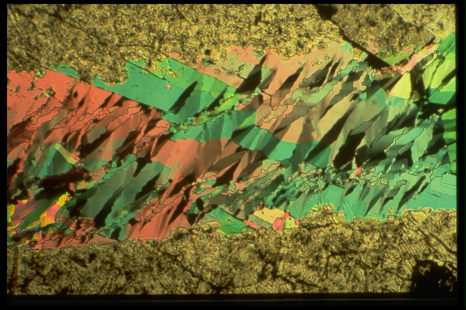
Kinking and Rotation Recrystallization in DCB. This series shows room temperature, dextral shearing of triclinic C6H4Cl2, para-dichlorobenzene (DCB). The magnification is lower than for previous slides. The width of the field of view is now about 2mm. The shear strain rate was about 1% per minute. |
|
| 22. The brown areas at the top and bottom of the
picture mark the positions of the two frosted grips. The DCB in these
areas is brown because it is thicker at the grips than in the
unfrosted, window area between the grips. At this stage, the sample has already been sheared a lot and there are numerous left-dipping kink bands crossing the originally right-dipping, pink and green DCB laths. The active slip plane in DCB is parallel to the lath boundaries — like the basal slip plane in micas. The shearing displacement across the sample can be followed in this series of five slides by noting the changing relative positions of the large green patch at (35,20) relative to the small green patch at (50,50). Some of the kink bands have straight boundaries but many have markedly irregular boundaries. This results from MIGRATION of originally straight kink boundaries through the material. The kink at (63,20) can be watched as an example of what is going on here. At this stage, the kink has only moderately irregular boundaries and contains several subgrains separated by sub-horizontal boundaries. |
|
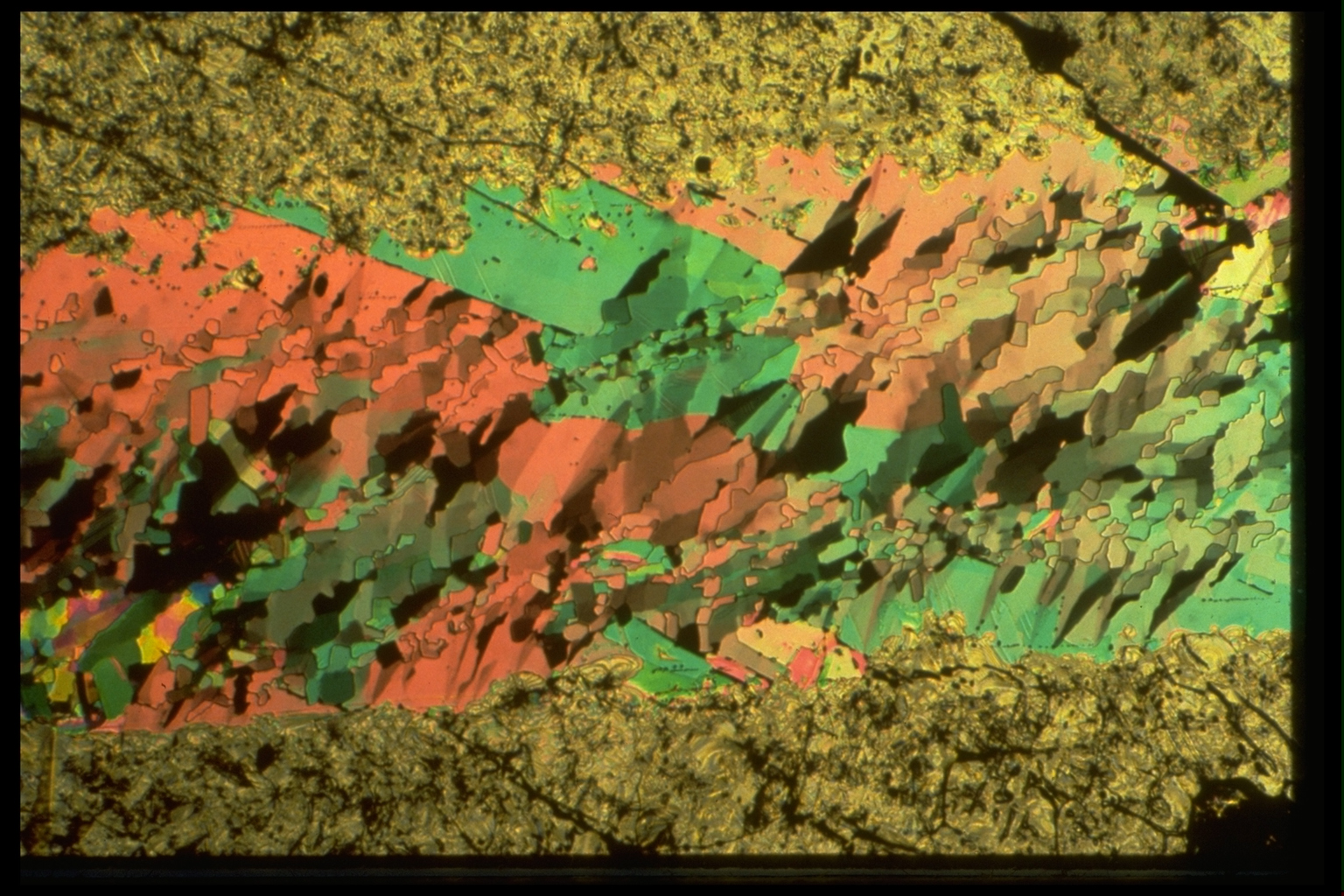
| 23. The kink described above, now at (72,20) has more irregular boundaries and better definition of its internal subgrains. |
|
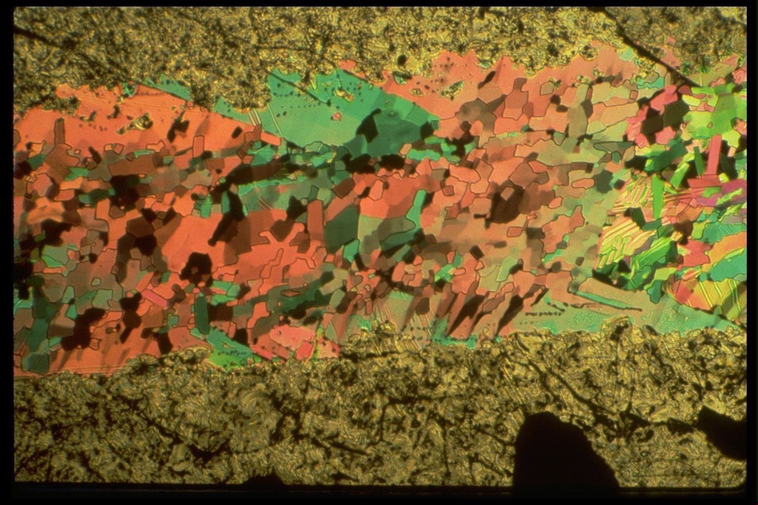
| 24. By this stage,
the original boundaries of the kink are no longer recognizable, but the
tan grain at (77,16) clearly represents the subgrain of similar color
at the top of the kink in the previous slide. This is a grain created
by kinking (to rotate its lattice strongly relative to the surrounding
pink material) and kink boundary migration (first to isolate the grain
from other parts of the kink, and then to allow it to grow). This is a
form of ROTATION RECRYSTALLIZATION. The other subgrains of the kink are
now gone, or evolved beyond recognition. Similar developments may be
seen elsewhere in the sample, especially near its margins. In the more
internal parts of the sample, the microstructural evolution is so rapid
that one cannot follow the histories of individual kinks or grains or
subgrains with confidence. The lamellar structures coming into the field of view around (90,45) are twin lamellae. |
|
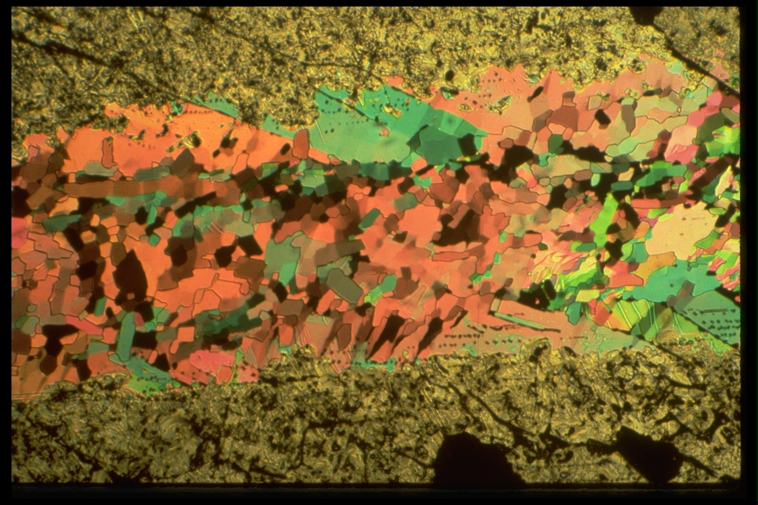
| 25. Around (87,43) one can see a kink band (grey) transverse to the twins. This is seen again, at (86,44) in the next image. More twins can be seen (in this image, 25) in the big green crystal around (33,16). Here, two twin lamellae are being eaten into by a brown grain from below. One can go backwards through the images to see that these twins are present in image 23 but are not yet developed in image 22. In image 26, they are almost entirely ingested by the growing brown grain. |
|
| 26. Notice the pink-brown RIBBON GRAIN that now
exists, parallel to the shear direction, centered at (20,25). Ribbon
quartz grains like this, with subgrain boundaries at a high angle to
the ribbon length, are common in deformed quartzo-feldspathic gneisses.
They are sometimes interpreted as stretched original grains. Does this
seem a likely explanation here? (ans) It is instructive to observe that the central part of this sample was once strongly kinked, despite the absence of obvious kinks now. Predict the origin of the small, elongate, nearly extinguished grain at (72,12). Then go back through the images to see whether you are right or wrong |
|